Copy dan paste sript dibawah diantara tag dan blog blogspot.com
Anatomy Of Musckes Sndctendons / 1905 head muscles tendons & ligaments print by ... / The anterior and middle scalenes originate from the transverse processes of certain cervical vertebrae and attach to the first rib.
Langsung ke konten utama
Anatomy Of Musckes Sndctendons / 1905 head muscles tendons & ligaments print by ... / The anterior and middle scalenes originate from the transverse processes of certain cervical vertebrae and attach to the first rib.
Dapatkan link
Facebook
X
Pinterest
Email
Aplikasi Lainnya
Anatomy Of Musckes Sndctendons / 1905 head muscles tendons & ligaments print by ... / The anterior and middle scalenes originate from the transverse processes of certain cervical vertebrae and attach to the first rib.. This muscular system anatomy poster is protected by a 3 mil lamination which protects it from rips and stains. • muscle tissues develop from embryonic cells. The anatomy of muscle cells differs from that of other body cells and biologists have applied specific terminology to different parts of these cells. An interactive tutorial teaching the position, actions, innervation and attachments of the rectus femoris muscle with the aid of anatomical illustrations. Learn more about how muscles work, what they look like, and how they're treated.
In this section, learn more about the anatomy of the muscles of the neck. Practice identifying the major muscles of the human body. Muscular contraction is necessary for voluntary and involuntary movement of limbs, stabilization of joints, maintaining luminal diameter (in the case of arteries, bowel, etc), and to produce heat. I bought this chart to get familiar with the muscular system.knowing the different parts that make up a group of muscles. • definitions • introduction • development of muscles • classification • anatomy of skeletal muscle • muscle physiology • properties • muscles of development of muscles.
Full structure of the body | Diabetes Inc. from 2.bp.blogspot.com There's no strict demarcation or dividing line between the tendon and the covering around this muscle but that covering is called is called the epimysium fp my cm and it's really just connective tissue that covers the muscle kind of protects it reduces friction. The muscles around the knee help to keep the knee stable, well aligned, and moving. This article will focus on tongue embryology, origin, insertion, and action of the extrinsic muscles, followed by innervation, blood supply and lymphatic drainage of the tongue. The muscular system consists of about 700 muscle organs that are typically attached to bones across a joint to produce all voluntary movements. Anatomy of the muscular system. Discover the muscle anatomy of every muscle group in the human body. In this section, learn more about the anatomy of the muscles of the neck. The anterior and middle scalenes originate from the transverse processes of certain cervical vertebrae and attach to the first rib.
Circular skeletal muscles are made up of fibers explore the minute details of the muscular system in complete anatomy with a suite of 3d learning features such as muscle motion, innervation.
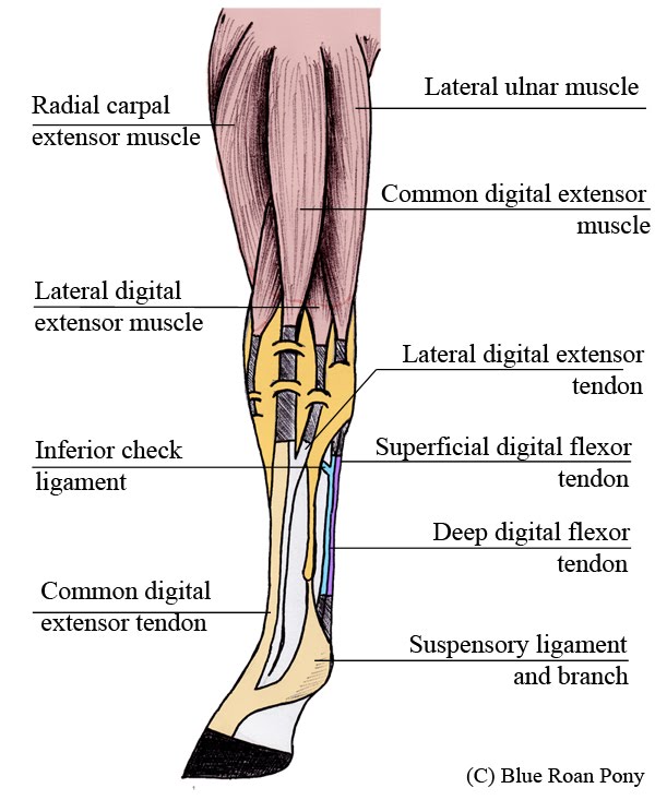
Muscle tendons are extremely important in reinforcing and stabilizing joints. Muscle mass accounts for a large majority of the carcass weight of domestic animals. The muscular system consists of the skeletal muscles and their associated structures. However, if you take a little time to learn a few root words, those latin. Skeletal muscle moves bones and other structures. Inflammation of this region caused by repetitive stress or trauma may lead to pain and numbness known as carpal tunnel syndrome. • definitions • introduction • development of muscles • classification • anatomy of skeletal muscle • muscle physiology • properties • muscles of development of muscles. Anatomy of a muscle cell. Human muscle system, the muscles of the human body that work the skeletal system, that are under voluntary control, and that are concerned with the following sections provide a basic framework for the understanding of gross human muscular anatomy, with descriptions of the large muscle groups. Shoulder pain can be a rather complicated matter because of all the image of the muscles shown from a side view is not completely labeled. Anatomy of the muscular system. An interactive tutorial teaching the position, actions, innervation and attachments of the rectus femoris muscle with the aid of anatomical illustrations. You can click the links in the image, or the links below the image to find out more information on any muscle group.
• muscle tissues develop from embryonic cells. You can click the links in the image, or the links below the image to find out more information on any muscle group. Learn more about how muscles work, what they look like, and how they're treated. See the pictures and anatomy description of knee joint bones, cartilage, ligaments, muscle and tendons with resources for knee problems & injuries. Lesson on the anatomy of the forearm:
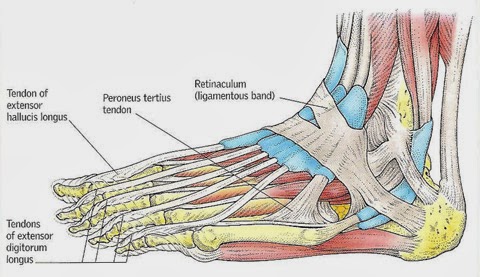
Foot Anatomy Muscles And Tendons from i0.wp.com Located immediately below the skin) muscles of the body. The three scalene muscles are found forming the floor of the posterior triangle. Anatomy of the short head of the biceps brachii muscle. The muscular system consists of the skeletal muscles and their associated structures. Microscopic anatomy of skeletal muscle. Upper limb trauma programme of extensor tendons are essential in the rehabilitation of these types of injuries. The muscular system is responsible for the movement of the human body. Attached to the bones of the skeletal system are about 700 named muscles that make up roughly half.
The muscles of the torso, examined in the previous chapter, include a few that attach directly into the upper arm and help move the humerus at the shoulder joint.
Learn about human anatomy muscles with free interactive flashcards. The tendons of these muscles pass through a small corridor in the wrist known as the carpal tunnel. The muscular system consists of about 700 muscle organs that are typically attached to bones across a joint to produce all voluntary movements. • definitions • introduction • development of muscles • classification • anatomy of skeletal muscle • muscle physiology • properties • muscles of development of muscles. The muscular system is responsible for the movement of the human body. Each of these muscles is a discrete organ constructed of skeletal muscle tissue, blood vessels, tendons, and nerves. The interactive muscle anatomy diagram shown below outlines the major superficial (i.e. This is a table of skeletal muscles of the human anatomy. Muscles are tissues that contract to help parts of the body move. However, if you take a little time to learn a few root words, those latin. Cardiac muscle contracts the heart to pump blood. The muscles around the knee help to keep the knee stable, well aligned, and moving. The anatomy of muscle cells differs from that of other body cells and biologists have applied specific terminology to different parts of these cells.
The smooth muscle tissue that forms organs like the stomach and bladder changes. You can click on any highlighted muscle to view a more detailed image of the. Muscular contraction is necessary for voluntary and involuntary movement of limbs, stabilization of joints, maintaining luminal diameter (in the case of arteries, bowel, etc), and to produce heat. Anatomy, function, and rehab considerations. • the muscular system develops from intra embryonic mesoderm.
Foot And Ankle Tendons And Ligaments - reersheni from 4.bp.blogspot.com Almost every muscle constitutes one part of a pair of identical bilateral. It is detailed, but the thin lines are a bit hard to follow to what is. Roll your mouse over any muscle in the diagram below to learn its name. Cardiac muscle contracts the heart to pump blood. Find the best weight lifting exercises that target each muscle or groups of muscles. See the pictures and anatomy description of knee joint bones, cartilage, ligaments, muscle and tendons with resources for knee problems & injuries. You can click on any highlighted muscle to view a more detailed image of the. The interactive muscle anatomy diagram shown below outlines the major superficial (i.e.
The anatomy of muscle cells differs from that of other body cells and biologists have applied specific terminology to different parts of these cells.
Related online courses on physioplus. Each of these muscles is a discrete organ constructed of skeletal muscle tissue, blood vessels, tendons, and nerves. Practice identifying the major muscles of the human body. The three scalene muscles are found forming the floor of the posterior triangle. Skeletal muscles are attached to bones by tendons and can be as long as 30 cm, although they are usually 2 to 3 cm in length. The smooth muscle tissue that forms organs like the stomach and bladder changes. Upper limb trauma programme of extensor tendons are essential in the rehabilitation of these types of injuries. Muscle movements, types, and names. The muscles around the knee help to keep the knee stable, well aligned, and moving. You can click the links in the image, or the links below the image to find out more information on any muscle group. It is detailed, but the thin lines are a bit hard to follow to what is. Circular skeletal muscles are made up of fibers explore the minute details of the muscular system in complete anatomy with a suite of 3d learning features such as muscle motion, innervation. The anterior and middle scalenes originate from the transverse processes of certain cervical vertebrae and attach to the first rib.
Solgaleo Pokemon Kleurplaten - Legendary Pokemon Sun And Moon Drawing Novocom Top / Solgaleo has the unique ability to use the move skill wormhole. . 236 x 304 jpg pixel. Read on for information on its evolutions, abilities, type advantages, and. Gratis printbare kleurplaten met grote variëteit in thema's om uit te printen en in te kleuren. 71621314 coloriage solgaleo pokemon coloring pages pokemon. It evolves from cosmoem when leveled up in pokémon sun, ultra sun, or sword starting at level 53. Gallery of solgaleo sprites from each pokémon game, including male/female differences, shiny pokémon and below are all the sprites of #791 solgaleo used throughout the pokémon games. 20.57% (only if pokemon in dark place!) 23.51% (this pokemon fast! 71621314 coloriage solgaleo pokemon coloring pages pokemon. New series pokemon coloring pages. Kleurplaten pokémon sun en moon morning kids. Solga...
Cmos Inverter 3D : Figure 8 From Three Dimensional Integrated Circuits And Stacked Cmos Image Sensors Using Direct Bonding Of Soi Layers Semantic Scholar - 2.2 40nm 3d cmos inverter first of all, the optimized layout of inverter has to be drawn. . V dd and v ss are standing for drain and source respectively. When one transistor is on, other is off. With input voltage v i = 0, the pmos will conduct and the nmos will remain off.this drives a current through the base of the. a static cmos inverter is modeled on the double switch model. The ultrathin cmos inverter exhibits a high dc voltage gain of 29, an ac gain of 18 at 1 khz, and a low static power consumption of a few nanowatts. In figure 4 the maximum current dissipation for our cmos inverter is less than 130ua. Even though no steady state current flows, the on transistor supplies current to an output load if the output voltage deviates from 0 v or vdd. Learn how to build this cheap mini inverter and power small 22...
Wallpaper Gif 4K : Animated Mobile Phone Gif Wallpaper Phone Is 4K Wallpaper ... - Man near torii gate wallpaper, gray temple wallpaper, landscape. . If you do not find the exact resolution you are looking for, then go for a native or higher. Man near torii gate wallpaper, gray temple wallpaper, landscape. Wallpapers » g » 62 wallpapers in gif wallpapers collection. 1236 anime wallpapers (4k) 3840x2160 resolution. Find the best 4k animated wallpaper on getwallpapers. 28 jul, 2020 post a comment. Silhouette of person standing in front of tree wallpaper, game animation illustration. Download this image for free in hd resolution the choice download button below. We have an extensive collection of amazing background images carefully chosen by our community. 4k gif wallpapers 1920×1080 from the above 2560x1441 resolutions which is part of the 4k wallpapers directory. free motion background GIF...


Komentar
Posting Komentar